Perioperative hypothermia
Sarah E. Allen, DVM, DACVECC
Massachusetts Veterinary Referral Hospital, Woburn, MA
NORMAL THERMOREGULATION
Body temperature is closely maintained in mammals around an optimal set point at which ideal cellular function can occur. This is a complex process that ultimately results in a balance between heat production and heat loss. The hypothalamus acts as the main regulator of this process with multiple sensors throughout the body – in the skin, thoracic and abdominal viscera and spinal cord.
The body can be viewed as two thermal compartments: the core and peripheral compartments. The core compartment is well perfused with a mostly constant temperature. The peripheral compartment is composed of the extremities. The peripheral temperature is dependent on heat transmission from the core via blood flow and heat conduction from adjacent tissues.
Heat is generated by metabolism and muscle activity. The brain, heart and abdominal organs are the main generators of metabolic heat. The basal metabolic rate is increased by elevations in thyroxine, and increased sympathetic stimulation with release of epinephrine and norepinephrine. Increased muscular activity in the form of shivering is also used as a means of generating heat. Heat that is produced by the body is retained to allow maintenance, or an increase in the core body temperature. Heat retention occurs by physiologic responses including piloerection and peripheral vasoconstriction, as well as behaviors such as seeking shelter and warmth, and curling up.
Hypothermia
Hypothermia is defined as body temperature of less than 38°C (100.4°F). Hypothermia is classified by degree of severity as:
- Mild: 32-37°C/ 89.6- 98.6°F
- Moderate: 28-32°C/ 82.4-89.6°F
- Severe: <28°C/ 82.4°F
With the initiation of hypothermia, compensatory responses are stimulated. At a body temperature of < 34°C (93.2°F), vasodilation and decreased metabolic rate occur. Below 31°C (87.8°F), thermoregulation ceases completely.
Primary hypothermia is due to prolonged exposure to low environmental temperatures, where the processes of heat production and retention are overwhelmed. Some patients may be more vulnerable to environment-induced hypothermia due to decreased body condition, increased body surface area or a decreased ability to generate heat. Neonates, geriatrics and patients suffering from chronic illnesses that result in decreased body fat and muscle may be more susceptible to environmental hypothermia.
Secondary hypothermia is a result of disease, surgery, or other processes that cause alterations in heat production, heat retention or thermoregulation. Clinical signs of secondary hypothermia are more pronounced at higher temperatures and may vary from the temperature ranges defined in primary hypothermia. A common cause of secondary hypothermia in small animal patients is anesthesia. Perioperative hypothermia consists of 3 phases, starting with an initial rapid decrease in the core temperature of 1-1.5°C in the first hour. Anesthetic medications cause vasodilation which results in redistribution of heat from the core to the periphery. This is accompanied by a decrease in the temperature threshold needed to cause reflex vasoconstriction. This phase is followed by a slower decrease in temperature over the next 2-3 hours as heat loss exceeds the rate of metabolic heat production. Metabolic heat production is decreased by 20-30% by anesthetic medications. During surgery there is increased heat loss by all means of heat transfer. The plateau phase is the final phase where the metabolic heat production is equivalent to continued heat loss. At this point the core temperature has decreased to a point that vasoconstriction occurs and heat loss is decreased.
The consequences of primary and secondary hypothermia are similar, although it is believed that patients with secondary hypothermia are more symptomatic at higher temperatures. Hypothermia has many physiologic effects and consequences. In the anesthetic setting, postoperative hypothermia has been associated with increased mortality in humans. A major side effect of hypothermia is a decrease in the metabolic rate. This includes decreased hepatic metabolism, which can extend the duration of effects of anesthetic medications and result in prolonged hypothermia. In the kidneys, hypothermia initiates a cold diuresis due to increased glomerular filtration rate (GFR) and decreased sensitivity to antidiuretic hormone (ADH), which can result in hypovolemia. With more severe hypothermia, renal effects can progress to decreased renal blood flow, decreased GFR and ischemic renal tubular damage, which can lead to acute renal tubular necrosis. Hypothermia can result in hypocoagulation due to thrombocytopenia, platelet dysfunction, and alterations in coagulation factor functions. Cardiovascular consequences of hypothermia include an initial catecholamine stimulated increase in heart rate and blood pressure, which increases myocardial oxygen demand and consumption. Hypothermia induces a left shift in the oxygen-hemoglobin dissociation curve with decreased oxygen release to the tissues. Combined with reflex vasoconstriction, this results in significant tissue hypoxia, especially to the skin and extremities. With more severe hypothermia there is decreased catecholamine responsiveness, resulting in bradycardia and hypotension. Arrhythmias can occur due to myocardial ischemia. Ventricular fibrillation, due to myocardial irritation is a commonly documented rhythm in patients with severe hypothermia. Hypoxia is propagated by a decrease in respiratory rate and tidal volume. Prolonged recovery from anesthesia due to hypothermia and deceased drug metabolism can cause altered mentation. Hypothermia has also been documented to cause altered and suppressed function of the immune system, resulting in increased surgical site infections, and altered wound healing in human and veterinary patients.
Treatment
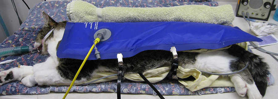
The main difference between the treatment of primary and secondary hypothermia is that the best treatment for secondary hypothermia is prevention. Prevention of perianesthetic hypothermia requires close patient monitoring with rectal or esophageal thermometers. Indwelling probes allow for continuous temperature monitoring. Therapies to prevent and treat hypothermia are very similar. Heat loss can be prevented with passive surface rewarming in the form of a blanket covering the patient, and between the patient and conducting surfaces (exam/surgery tables). Extremities can be wrapped in insulating material such as bubble wrap. Active surface rewarming works to increase the temperature of the air and surfaces surrounding the patient, and to decrease heat loss. Forced warm air blankets and circulating water blankets can significantly decrease heat loss. Active core rewarming acts to increase the temperature of the core by introducing heat centrally. This is most commonly done in the form of warm IV fluids and inhalation of warm, humidified air. More aggressive forms of core rewarming include warm pleural and peritoneal lavage, warm water enemas, and warm saline bladder lavage. Warmed IV fluids are helpful in preventing hypothermia but are of minimal assistance in rewarming hypothermic patients. IV fluids can be warmed to 40°C (104°F) via microwave or incubator. An in-line fluid warmer helps to maintain the temperature as the fluid is being administered. Peritoneal and pleural lavage fluids should be warmed to 40-43°C and can assist in rewarming, and preventing further heat loss in surgical patients.
In rewarming hypothermic patients, all of the previously mentioned preventative measures can be employed based on the severity of the hypothermia. With mild hypothermia (>32°C/ 89.6°F) passive surface rewarming may be sufficient. With more severe hypothermia (>28°C/ 82.4°F) active external rewarming is required with the main efforts aimed toward the core region rather than the extremities. With severe hypothermia (<28°C/ 82.4°F) active core rewarming is mandatory. In non-surgical patients, peritoneal and pleural lavage can be performed. Warm lavage allows heat transfer from the fluids to the bowel and heart, and can help prevent further decrease in temperature. Heat transfer is able to occur as long as the lavage fluid temperature exceeds the core temperature, ideally 40-43°C (104-109°F). A peritoneal catheter or chest tube can be placed with sterile technique if not already in place; 10-20 ml/ kg exchanges of warm lavage can be performed with dwell time effecting extent of heat transfer. Transfers can be repeated as needed to assist in regulating body temperature. Similarly, warm lavage of the urinary bladder and colon is also an option of unknown benefit due to limited surface area, but is less invasive than placing catheters accessing the peritoneal and thoracic cavities. Many hypothermic patients willrequire cautious IV fluid resuscitation to assist in restoring blood pressure and effective circulating volume. As hypothermia progresses, a decreased affinity for catecholamines leads to decreased cardiac contractility and vasodilation. This may result in minimal response to IV fluid therapy and should not automatically indicate a need to give excessive volumes of fluids in the absence of evidence of ongoing hypovolemia. As the temperature returns to normal, rebound vasoconstriction and increased contractility may result in fluid overload. Warm IV fluids are recommended so that fluid resuscitation does not potentiate the hypothermia. The ideal rate at which to rewarm is unknown, but a general recommendation is 1-2°C per hour. To prevent hyperthermia, active rewarming efforts are stopped once the patient has reached 37°C (100.4°F).
Rewarming complications can affect the prognosis of hypothermic patients. With rewarming, there is an increase in the metabolic rate and oxygen consumption that may not be appropriately supported in patients that are still hypotensive or hypoventilating. Supplemental oxygen may be beneficial in preventing hypoxia from this increase in oxygen demand. With external warming, a condition known as afterdrop can occur. In this case, the core temperature continues to decrease due to peripheral vasodilation and movement of warm core blood to the periphery, and return of cold blood to the core. Afterdrop may be prevented by instituting core rewarming in addition to peripheral, and by directing peripheral rewarming attempts at the trunk and not the limbs. Another complication concern is rewarming shock, when there is rapid vasodilation from external warming, which results in significant venous blood pooling and circulatory collapse. Injuries due to active rewarming efforts can occur in the form of thermal burns if there is excessive heat used (>42°C/ 107.6°F), prolonged contact time of the patient to heat, or a lack of insulation between the heat source and patient.