Feline head imaging: Continuity with practitioner and radiology
Nedra Wilson, BVetMed, DACVR, MRCVS
Ethos Veterinary Health
Posted on 2016-11-08
Diseases of the head are often initially imaged with radiographs in the clinic. Based on the findings from these radiographic studies, our patients may need immediate intervention or advanced imaging (CT or MRI) to obtain the necessary information that we as clinicians can use to help advise owners in making good decisions.
Head imaging is challenging
Skull radiography has long been difficult in any setting. Cats are typically stressed and not used to being transported from their home environments. Optimal positioning, standardization of views and variations in the radiography system can all present different artifacts that make interpretation difficult. In the feline, the main indications for skull radiography include trauma, nasal discharge (acute, chronic, epistaxis), facial deformation and dentistry.
One of the most important challenges with skull radiographs is optimal positioning. A lot of time feline patients present with cardiac, renal and hepatic disease, which requires special consideration with sedation or anesthesia requirements. Although less convenient, general anesthesia is most ideal for all feline patients that require head imaging. We may also consider that if a cat needs head imaging with general anesthesia that the gold standard examination, such as CT, may be recommended from initiation of the problem.1 However, it is acknowledged that most owners want to start simple, then move to more advanced things as information is presented to them.
The jury is still out: feline skull radiographs may or may not be helpful
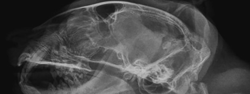
The feline skull has very complex anatomy with more than fifty bones that are usually paired. This makes imaging difficult because of superimposition of many of these structures.2 Most of these bones cannot be imaged on radiographs because they fuse. However, with well-positioned radiographs that have good detail and contrast, meaningful information may be obtained. Standard views should always include a lateral and a DV (or VD). Special views to see the tympanic bullae and temporomandibular joints may also be obtained.2 However, lateral oblique views of the maxilla and mandible tend to be of little benefit when the patient is not well positioned and the images are labeled inappropriately. Time can be wasted in obtaining the radiographs, more anesthesia time is required and there is increased exposure to personnel taking them.
A review of skull anatomy
Larger bones of the skull that may be evaluated on radiographs include the incisive, nasal, maxillary, lacrimal, frontal, zygomatic, pterygoid, sphenoid, parietal, temporal and occipital.3 The nostrils are rostral openings to the nasal cavity, extending caudally to the cribriform plate and nasopharynx.4 The maxillary bone houses the nasal cavity, along with the smaller incisive and nasal bones. Ventrolaterally are the paired maxillary recesses that are blind-ended and small in the feline. Within the rostral nasal cavity are nasal turbinates, which are thin scrolls of bone that make up the dorsal and ventral nasal conchae. In the feline, the ventral conchae are short but very thick.4 The nasal turbinates obstruct airflow, while the nasal passages are what allow airflow. There are four nasal passages, which are the dorsal, middle, ventral and common meatus. The common nasal meatus is what communicates with all three of the other meati, with its medial margin being the nasal septum and may be curved in the cat. The caudal portion of the ventral meatus opens to the nasopharyngeal meatus, which communicates with the oropharynx. The paranasal sinuses also communicate with the nasal cavities.
The caudal nasal cavity houses the ethmoid turbinates that attach to the cribriform plate. In the feline, the ethmoid turbinates are well developed and invade the lower part of the frontal sinuses.3 In comparison to the canine, the feline frontal sinuses are not compartmentalized and male cats have larger frontal sinuses which is the reason for their larger heads.
Common diseases of the feline head
In cats, nasal neoplasia and chronic sinorhinopathies account for approximately 70% of chronic nasal disease.4 These two manifestations can be difficult to differentiate on radiographs and tend to have similar characteristics clinically,5 as well as on imaging that is initiated to help with a diagnosis. On radiographs, increased soft tissue opacification of the nasal cavities and paranasal sinuses, along with bone lysis of turbinates and paranasal sinuses may be seen in both conditions.4 In cats, characteristics most seen with neoplasia include unilateral soft tissue opacification of the nasal cavity and paranasal bone lysis with tooth loss.6
An important paper to be aware of when imaging the feline head
A recent paper published by colleagues at Cornell University School of Veterinary Medicine documents a serious concern with the use of mouth gags in cats.7 The Cornell group used computed tomography (CT) and nonselective digital subtraction angiography to show that in cats where mouth gags are used, for example in dental procedures or for skull radiographs, it is possible to reduce maxillary arterial blood supply to the brain. The maxillary arteries are the main blood supply to the Circle of Willis. They demonstrate that when there is a reduction in blood flow to the brain, it is possible for collateral blood flow to support the brainstem and cerebellum, but this may not be sufficient to supply the entire cerebrum. Therefore, based on their findings, some cats may develop bilateral blindness and cerebral ischemia as a result of using a mouth gag. While there are multiple factors that likely go into this process, the fact that it is possible may change how mouth gags are used in our feline patients.
Cross sectional imaging is all that
One of the better ways to image the feline head is with computed tomography, which can give cross sectional and three-dimensional views through the anatomy of interest. Information may be obtained without superimposition of structures and therefore greater detail of anatomy. The use and availability of CT has grown rapidly in human medicine, which has spilled over into veterinary medicine, where many referral practices are able to perform CT examinations and generate a report from a radiologist. Although general anesthesia is required for imaging our feline patient heads, the total time needed for acquisition of the study may be less overall than what may be required to obtain useful radiographic views.
References
- Lasonsky J, Abbot L, Kuriashkin I: Computed Tomography of the Normal Feline Nasal Cavity and Paranasal Sinuses, Vet Radiol Ultrasound, Vol 38: pp 251-258, 1997.
- Hammon G, Sullivan M, Weinrauch S, King A: A Comparison of the Rostrocaudal Open Mouth and Rostro 10° Ventro-Caudodorsal Oblique Radiographic Views for Imaging Fluid in the Feline Tympanic Bulla, Vet Radiol Ultrasound, Vol 46: pp 205-209, 2005.
- Thrall D, Robertson, I: Atlas of Normal Radiographic Anatomy & Anatomic Variants in the Dog and Cat, ed 1, St. Louis, 2011, Saunders.
- Saunders J, Schwarz T: Nasal Cavities and Frontal Sinuses. In Schwarz T, Saunders J, editors: Veterinary Computed Tomography, ed 1, UK, 2011, John Wiley & Sons Ltd.
- Shanaman M, Seiler G, Holt D: Prevalence of Clinical and Subclinical Middle Ear Disease in Cats undergoing Computed Tomographic Scans of the Head, Vet Radiol Ultrasound, Vol 53: pp 76-79, 2012.
- Tromblee T, Jones J, Etue A, Forrester D: Association Between Clinical Characteristics, Computed Tomography Characteristics, and Histologic Diagnosis for Cats with Sinonasal Disesase, Vet Radiol Ultrasound, Vol 47: pp 241-248, 2006.
- Scrivani P, Martin-Flores M, Van Hatten R, Bezuidenhout A: Structural and Functional Changes Relevant to Maxillary Arterial Flow Observed During Computed Tomography and Nonselective Digital Subtraction Angiography in Cats with the Mouth Closed and Opened, Vet Radiol Ultrasound, Vol 55: pp 263-271, 2014.
Further reading
- Fischetti A, Gisselman K, Peterson M: CT and MRI evaluation of Skull Bones and Soft Tissue in Six Cats with Presumed Acromegaly Versus 12 Unaffected Cats, Vet Radiol Ultrasound, Vol 53: pp 535-539, 2012.
- Karnik K, Reichle J, Fischetti A, Goggin J: Computed Tomography Findings of Fungal Rhinitis and Sinusitis in Cats, Vet Radiol Ultrasound, Vol 50: pp 65-68, 2009.
- Oliveira C, O’Brien R, Matheson J, Carrera I: Computed Tomography Features of Feline Nasophyarngeal Polyps, Vet Radiol Ultrasound, Vol 53: pp 406-411, 2012.
About the author
Dr. Wilson received her Doctor of Veterinary Medicine (BVetMed, Merit Finals part III) from the Royal Veterinary College, University of London, UK in 2006. She returned from across the pond and completed a one-year rotating small animal medicine and surgery internship at a private practice in Massachusetts, followed by working two years as a staff emergency clinician at DoveLewis Emergency Animal Hospital in Portland, OR. Her three-year residency in Diagnostic Imaging from Cornell University was completed in July 2012. She was subsequently awarded a clinical fellowship for one year with a joint appointment in the Department of Biomedical Sciences at Cornell University School of Veterinary Medicine and the Imaging department at Cornell University Hospital for Animals. For the past several years, she has worked in private practice in New Hampshire before joining the IVG network in October 2015. Dr. Wilson became board certified by the American College of Veterinary Radiology in September 2014. She is also a current member of the Royal College of Veterinary Surgeons (MRCVS). Dr. Wilson has strong professional interests in computed tomography (CT), musculoskeletal ultrasound and imaging of exotics/marine mammals. |