Radiography vs. ultrasound in the dog with acute abdominal signs
Miriam M. Shanaman, VMD, MS, DACVR
Veterinary Specialty Hospital, San Diego, CA
Posted on 2016-09-20
In the emergency setting, the primary goal of diagnostic imaging is to help differentiate surgical from non-surgical conditions. The benefits and limitations of survey radiography and abdominal ultrasound are outlined below.
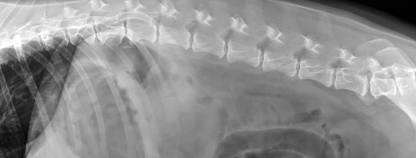
Survey Radiography
One of the primary benefits of survey radiography is improved sensitivity for the detection of pneumoperitoneum. In the absence of recent laparotomy, peritoneal free gas is considered a surgical condition with differential diagnoses including the following: ruptured hollow viscus, ruptured abscess or necrotic neoplasm, and penetrating wound.
In cases of small bowel obstruction, the goal of survey radiography is to identify or rule out an obstructive bowel pattern. This requires the identification of two distinct populations of small bowel; one population that is normal in dimension and one population that is abnormally dilated. Accurate radiographic assessment of small bowel obstruction, therefore, requires the ability to unequivocally differentiate small from large bowel. Furthermore, if abnormal dilation is limited to the duodenum, a definitive diagnosis of mechanical obstruction may continue to be problematic as acute pancreatitis may result in a similar finding. Finally, if all of the small bowel is considered abnormally dilated, while distal small bowel mechanical obstruction remains possible, functional ileus remains a differential. Linear foreign bodies are particularly problematic as these may not present with the typical “two populations of bowel” described above. Instead, findings such as bowel plication, bunching, eccentric luminal gas bubbles, and gastric fluid distention (if a concurrent pyloric outflow tract obstruction is present) may be seen. Three-view abdominal radiographs (right lateral, left lateral and VD projections) are recommended in all vomiting patients. The left lateral view is particularly helpful for assessment of the pyloric outflow tract and proximal duodenum such that foreign bodies or mass lesions that may be obscured normally on the right lateral view may be outlined by gas and therefore visible on the left lateral view.
The primary non-GI etiology for acute abdominal signs in canine patients is acute pancreatitis. If there is sufficient inflammation, potential radiographic findings include decreased serosal detail, displacement of the proximal descending duodenum to the right and pylorus to the left, regional mass effect, duodenal and/or gastric fluid distention and caudal displacement of the transverse colon. These findings, however, are non-specific and do not permit differentiation of inflammatory from neoplastic pancreatic disease nor from other potential regional causes of similar imaging findings (such as infiltrative neoplasia of the stomach or proximal duodenum for example).
Abdominal Ultrasound
While abdominal ultrasound has reduced sensitivity for the detection of pneumoperitoneum, there is improved detection of small amounts of peritoneal free fluid. Sampling of peritoneal free fluid is always recommended to be performed with ultrasound guidance and may yield a surgical diagnosis (e.g., uroabdomen, non-traumatic hemoabdomen in the absence of coagulopathy, bile peritonitis, septic peritonitis).
In cases of suspect small intestinal or gastric outflow obstruction, the primary goal of abdominal ultrasound is to identify the distinct zone of transition between normal and abnormal bowel or to evaluate the pyloric outflow tract respectively. This requires a high level of expertise as long segments of bowel must be carefully scanned in patients that are often painful or vomiting. Furthermore, the sonographer must frequently evaluate the patients in variable recumbency as the visibility of certain bowel segments and portions of the stomach may vary accordingly. These types of examinations are often time-consuming, however, in the hands of an experienced sonographer will eliminate ambiguities regarding mechanical vs. functional conditions of the bowel in addition to the exact etiology for mechanical obstruction if present. In our experience, bowel plication secondary to linear foreign material is much more evident sonographically than on survey radiographs; especially when the foreign body remains limited to the proximal duodenum.
Ultrasound is used routinely in the identification of pancreatitis and permits staging of disease such that potential complications including extra-hepatic biliary tract obstruction, abscess/ pseudocyst formation, duodenitis, gastritis and portal vein thrombosis may also be identified. Furthermore, in cases of suspect pancreatic neoplasia, ultrasound permits the guided sampling of mass lesions and regional enlarged lymph nodes or ascites if present.
Conclusion
Ultimately, initial survey radiography followed by abdominal ultrasound is optimal given the complimentary nature of these modalities.
About the author
|
 A diplomate of the American College of Veterinary Radiology, Dr. Miriam Shanaman joined VSH in August 2013. She completed both her undergraduate and veterinary training at the University of Pennsylvania, earning her VMD in 2009. Following graduation from veterinary school, Dr. Shanaman completed a rotating internship in small animal medicine and surgery at New York City Veterinary Specialists in New York, NY. Immediately following her internship, Dr. Shanaman completed a 3-year combined Master’s degree and clinical residency in diagnostic imaging at the University of Illinois, earning her board certification in 2013. Through her Master’s research and clinical training, Dr. Shanaman has published multiple articles addressing the use of contrast-enhanced multi-detector CT in the assessment of awake or minimally sedated canine patients with acute abdominal signs. Dr. Shanaman also has extensive experience with the use of contrast-enhanced ultrasound in the same context. Her clinical interests include evaluating the utility and efficacy of multi-detector CT in the emergency setting to assess conditions of the respiratory, gastrointestinal and urinary tracts in addition to global assessment of patients suffering from polytrauma.

