Wet and wild: Aquatic animal anesthesia basics
Stephen Cital RVT, SRA, RLAT
United Veterinary Specialty and Emergency, Oakland and San Francisco Zoo
For more visit www.stephencital.com
This blog touches on the basics of fish and amphibian general anesthesia. Species anatomy and physiology will be lightly covered, along with considerations when choosing a general anesthetic agent.
Basic anatomy & physiology
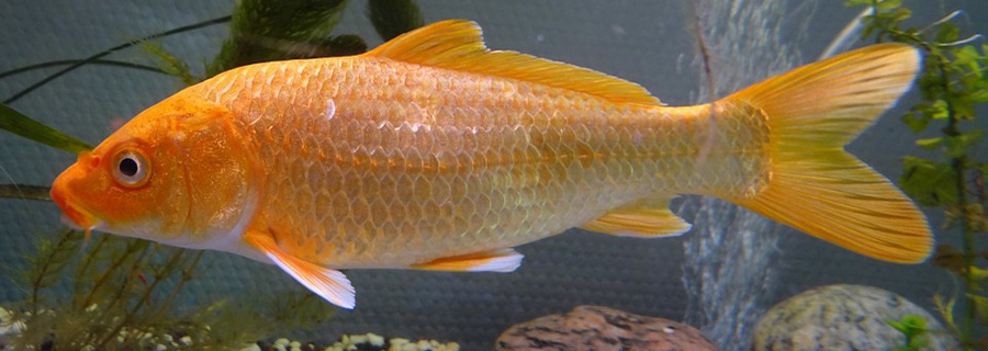
Fish represent one of the first vertebrate species on the evolutionary tree. The anatomy of fish, although similar to other vertebrates, clearly shows the progression of evolution through the ages. Fish embryos play a particularly interesting role in human embryotic studies since early fish embryos are nearly identical to early human embryos. Since fish have only a two-chambered heart (depending on what one considers a true chamber) and a gill system instead of an air- breathing respiratory system like that starting with more evolved air breathing fish species, then progressing to amphibians, make fish a particularly interesting taxa to work with and anesthetize. Fish are also becoming a more and more prized pet with owners willing to lay down the cash to treat, especially if they are a rare or exotic fish.
Amphibians have many similarities to reptiles. Like reptiles amphibians have a three-chambered heart and a whole separate lymph system with “lymph hearts”. Lymph is responsible for carrying substances absorbed through the skin into the body and is eliminated through the renal system. There are species-specific variations in septal fenestrations allowing mixing of deoxygenated and oxygenated blood like seen in some reptiles.
Amphibians, like reptiles, also have a renal portal system that can affect drug concentration levels between the cranial half of the body to the caudal half of the body. Injecting in the caudal half of the animal may lead to quicker excretion without a systemic effect.
Another fascinating physiological advantage that some amphibians have are adaptations for dealing with super cold environments. Already able to tolerate anoxic conditions well with the ability to slow their metabolism and switch to anaerobic metabolism, amphibians also have special proteins to fair long anorexic periods and ice. Ice nucleating proteins help keep ice crystals from forming intracellularly while glucose transporting proteins can move necessary glucose to vital organs during hibernation -like states. Amphibians also have a well-defined fibrinogen response to cold and have an active heat shock protein function to deal with varying temperatures.
The respiratory system is particularly interesting in amphibians. Many species go through a complete respiratory metamorphosis, from gills in the larval stage to lungs in the adult animals with a species variable cutaneous and buccopharyngeal respiration system. The highly variable respiratory systems in amphibians leave some species with external gills in neotenic species. It is notable to investigate thoroughly the primary mode of gas exchange in a particular species during its particular life stage for appropriate anesthetic agent administration.
- Branchial respiration: Use of gills for oxygen absorption from dissolved gasses in the water
- Cutaneous respiration: Gas exchange through the skin via superficial capillaries. This method is similar to passive dissolved gas exchange used with gills.
- Buccopharyngeal respiration: Oxygen absorption through the buccal cavity via a blood rich membrane
- Pulmonic respiration: Gas exchange via lungs. Reptiles, birds and amphibians all have primary lung sacs rather than alveoli containing lungs seen in mammals. However, certain species of amphibians do have sacculations that contain some alveoli.
Temperature regulation & metabolism
Being ectotherms, amphibians and fish are highly reliant on us for their preferred optimal temperature. They depend largely on their ability to move to warmer and cooler areas as part of their thermoregulation. Amphibians that have chromatophores can even change their skin color via light sensitive proteins in their epidermal cells to better reflect solar energy. Amphibians in general control their body temperature via evaporation of fluid from their skin, as well as peripheral capillary vasoconstriction and vasodilation. Temperature along with humidity will greatly affect the patient’s metabolism and has a direct correlation to acid-base and blood gas composition.
Amphibians produce most of their energy from aerobic metabolism. However, during long dives or more strenuous activity, anaerobic metabolism can be utilized. Prior to and after an anesthetic event housing a patient with a mosaic of temperature zones is recommended. Fish should be recovered in the same temperature water as their home environment. Intraoperatively keeping the patient at the species preferred temperature and humidity is vital. Like reptiles, prolonged hypothermia can lead to slow recoveries and immune suppression. Higher temperatures have the benefit of immune enhancement.
Larger amphibians have a decreased caloric intake need compared to smaller species. It should be noted that ill or recovering from surgery amphibians, like many species have an increased metabolic rate (1.5-2 times increase). This means they require a higher caloric intake.
Water quality
Water quality is highly important to any aquatic species. De-chlorinated and non-salinized water are two of the more important water quality factors, unless the fish is a salt water species of course. Each species will require differing pH levels, anywhere from 6.5-9.5. The water should be clean either by filter or portion replacement. Oxygenated water is also a very important factor, in particular for hospitalized patients or animals requiring an anesthetic procedure. Oxygenating water can be achieved by surface disruption from an agitator or a bubbler at the bottom of the tank. When preparing water for an anesthetic event, using a bubbler with 100% O2 will ensure well oxygenated water. If at all possible, using water from the patient’s tank is recommended.
Anesthetic induction
Induction methods for amphibians and fish are still an evolving science. Once thought effective, hypothermic induction is now considered inappropriate for painful procedures, as full unconsciousness is not achieved.
Some sources cite Propofol™ IV, IO or intracoelomically as an option, but difficulties placing indwelling IV catheters or species tolerance can complicate this method. Other anesthetic protocols including the addition of Propofol™ to the water have also been documented with success. Alflaxalone has had variable success either transcutaneously or intramuscularly. Alflaxalone in certain fish studies, such as Koi, have been successful. Other parental methods and induction agents, such as ketamine, have been used IM, and even in the dorsal lymph sacs of amphibians, but little has been written to support these methods and a regular means of anesthetic maintenance. Other adverse effects, like a prolonged recovery, have been associated with ketamine anesthesia. Traditional inhalant anesthetics via the patient’s airways are theoretically possible in most amphibian species, but is generally reserved for large species of amphibians that can more readily be intubated. “Bubbling” anesthetic gasses in oxygenated water has been used in fish but can be costly and unsafe for staff due to environmental contamination.
The preferred method for anesthetic induction in amphibians takes advantage of their highly absorbent skin. Also known as “immersion anesthesia,” this same method can be used in fish as well for initial sedation and then maintenance. Once the fish is able to be handled, a tube with with circulating anesthetic water containing Tricaine Methanesulfonate (MS-222) or benzocaine can be placed in the mouth to pass over the gills. Clove oil was once a common anesthetic in fish, but has fallen out of favor. MS-222 is the more widely used aquatic animal anesthetic with the most supporting literature.
Immersion in a sodium channel blocker, such as MS-222, allows easier and less traumatic induction in amphibians and fish with a wide safety margin. Prior to mixing with water, the compound is a dry, white salt-like substance. When mixed with water it dissolves readily, producing a clear acidic bath. This acidic bath alone can cause trauma to delicate soft tissue, so it will need to be buffered prior to use. Buffering the solution with sodium phosphate to a normal physiological range for a particular species is the easiest method and safest by not having to worry about ratios of ionized to unionized MS-222. A non-buffered solution can lead to complicating iatrogenic metabolic acidosis. Sodium bicarbonate (baking soda) is also commonly used, but has more specific instructions to achieve a desired pH of 7-7.4. Too much sodium bicarbonate can lead to undesirable pH levels leaving more un-ionized MS-222. The fresh water used for the mixing of the solution will also be a key factor in achieving an appropriate pH level, making this method more challenging.
Adverse effects of MS-222 include apnea. Some scientists believe transdermal respiration occurs when patients become apneic under anesthesia, stressing shallow, well oxygenated circulating water during the procedure if possible, these aquatic species can drown! Induction time is usually 10-30 minutes.
Benzocaine at 0.01%-0.03% is recommended for adult amphibians. Lower doses are indicated for larval and paedomorphic amphibians. Apnea is also a concern with benzocaine immersion anesthesia. Multimodal approaches are also encouraged with the use of opioids IM to reduce the required anesthetic agents.
Placing any of the solutions mentioned above that is within the preferred species temperature range in a plastic bag with the patient will help alleviate self-induced trauma to the patient during any rash movements during the induction process. Every amphibian species and even patient will have a variable induction rate and necessary concentration depending on their life stage, size and any pre-existing condition. When performing immersion induction, keeping the patient’s head and nares above the water is crucial to prevent accidental drowning.
Note: Toads can take longer than frogs for induction due to their slower absorption of fluids through their skin.
After placing the patient in the solution, the patient will go through an agitation/excitement phase that is usually accompanied by slight erythema in amphibians. The patient will them begin to lose its righting reflex. At this point, the patient should become anesthetized within minutes depending on size, temperature, solution concentration and life stage. For fish and amphibians alike, mechanical stimulation by pinching the tail fin or toe can help assess the depths of the animal. The anesthetic effect is variable for all the previously mentioned variables, but one can expect anesthetic periods anywhere from 5-20 minutes after a 5-minute bath.
For fish specifically, a pump will need to be set up to continuously pass oxygen rich MS-222 solution over the gills via a tube placed in the mouth of the patient. It is recommended to have a couple different concentrations of MS-222 solution and a supply of non-anesthetic water to adjust anesthetic depth, also acting as a ventilator for the fish.
Isoflurane, as mentioned above, can be used in the gas form more easily for larger species of amphibians. Isoflurane can also be applied directly to the skin of the animal at around 0.007-0.015 mL/g. The inhalant liquid can also be mixed with a water-based gel and applied to the skin at slightly higher doses.
NOTE: Always have plenty of fresh water available for emergency rinsing and soaking in the event of anesthetic complications
Intubation and volatile anesthesia are possible, but less effective, in amphibians due to their alternative respiratory systems.
Monitoring and Maintenance
Fish and amphibian anesthetic monitoring can really test a practitioner’s skills. One will have to rely heavily on close observation and his senses for respiration rate and quality, as well as anesthetic depth. Cardiac function and rate can be assessed by placing a Doppler directly over the heart or well lubricated eye. Electrocardiograms can be taken on fish and amphibians in the same fashion as with mammals, using a three or four lead electrode setup. The author prefers using small gauge hypodermic needles or wire instead of clips that can damage the patient’s soft tissues and bones. A reflective SpO2 transducer can be placed in the cloaca or esophagus of either species. Pulse oximetry should not be used at a definitive diagnostic tool in any species other than humans as they are not calibrated to animal blood hemoglobin. Reflexes used to determine anesthetic depth in mammals can also be used in fish and amphibians with the exception of the palpebral reflex since most fish do not have eyelids. Some species may experience gulping like behavior under anesthesia. This reflex can be associated with breathing in some species and being “light” in others.
Recovery
When recovering the amphibian patient, many of the same precautions we take in other species apply. A shallow fresh water bath at the species preferred optimal temperature should always part of the recovery process to clear away any residual topical anesthetics.
Recovering fish requires immediate relocation to oxygenated water at the optimal temperature for the particular species. Some patients can benefit from a soft cradle until the fish is able to “right” itself after the anesthetic period. For larger fish, it also may be optimal to have a slight current in the recovery tank for continued ventilator support enabling water to pass over their gills. Pain management is always recommended in any species.
Further reading
- Grimm, K et al. (2015). Lumb & Jones’ veterinary anesthesia (5th ed.). Iowa: Wiley Blackwell.
- Longley, L., & Fiddes, M. (2008). Anaesthesia of exotic pets. Edinburgh: Elsevier Saunders.
- McMillan, M & Leece E. (2011). Immersion and branchial/transcutaneous irrigation anaesthesia with alfaxalone in a Mexican axolotl, JVAA.
- Minter, L et al. (2014). The efficacy of alfaxalone for immersion anesthesia in koi carp (Cyprinus carpio), JVAA.
- O’Malley, B. (2005). Clinical anatomy and physiology of exotic species: structure and function of mammals, birds, reptiles, and amphibians. Edinburgh: Elsevier Saunders.
- Ross, L & Ross, B. (2008). Anaesthetic and Sedative Techniques for Aquatic Animals. Oxford: Blackwell
- West, G. (2014). Zoo animal and wildlife immobilization and anesthesia. Ames: Blackwell Publishing.


Wow!! Great article – thank you!
Thanks for sharing this. My hospital really doesn’t have the opportunity to work with very many exotics. In all honesty I had never really thought about fish and anesthesia. Would it be possible to post a video of this procedure?