
Seeing between the lines
Jennifer Brisson, DVM, DACVR
Massachusetts Veterinary Referral Hospital, Woburn, MA
History
A 4-month-old female Labrador Retriever is presented for progressive lethargy and decreased appetite of 10 days duration. Her owners report that she vocalized while trying to stand this morning, and then would not get up. She has a history of treatment responsive infectious tracheobronchitis (kennel cough complex) and pneumonia at 10 weeks of age.
Physical exam
On presentation, she is quiet, reluctant to stand, has swollen carpi bilaterally with generalized pain on palpation of her limbs, and is febrile (103.2˚F). Her physical examination is otherwise unremarkable, with clear lungs on auscultation.
The following radiographs were obtained:
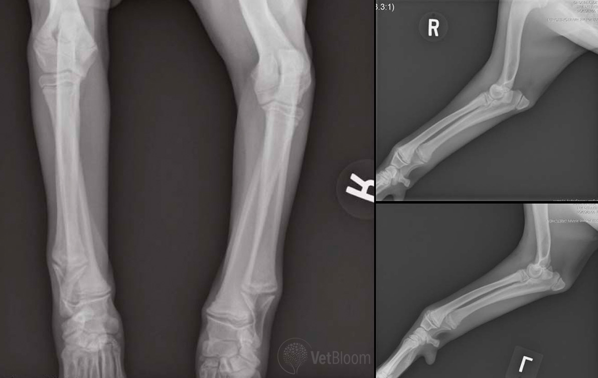
Radiographic diagnosis
Hypertrophic osteodystrophy
Hypertrophic osteodystrophy
Hypertrophic osteodystropy (HOD; a.k.a. metaphyseal osteopathy) is an idiopathic disease that affects young rapidly growing large and giant breed dogs. Osteodystrophy is a term used when a defect in bone formation occurs. HOD is reported in dogs between the ages of 2-8months, with no statistical sex predilection, though reports have noted males to be overrepresented. Great Danes, German Shepards, Weimeraners, Irish Setters, Boxers and Chesapeake Bay Retrievers are breeds reported with higher incidence. The diagnosis of HOD is made by physical examination and radiography.
Dogs with HOD may present for lameness, reluctance to walk, lethargy, anorexia, weight loss, and fever. On physical examination there may be firm, painful, warm swellings of the metaphyseal region of the long bones. The most common bones affected are the distal radius, ulna, and tibia. Other bones reported to have lesions include the femur, metacarpals/metatarsals, mandible, and scapula. The lesions are usually bilateral and symmetric.
Radiographic appearance
The radiographic findings in a patient with HOD consist of the “double physis sign.” This is linear metaphyseal sclerosis (increased opacity) and linear metaphyseal lysis (increased lucency) parallel to the physis, with perilesional tissue swelling. Necrosis, inflammation and hemorrhage at the proximal aspect of the physis and metaphysis causes osteolysis which manifests as a linear lucency. Parosteal cuffing, periosteal new bone, and soft tissue mineralization causes an increase in density adjacent to the metaphyseal osteolysis. The lesions are usually bilateral and symmetric.
Rarely, dogs with HOD develop periosteal new bone formation of the ventral mandible. This is similar in appearance to dogs with craniomandibular osteopathy, seen mostly in Terriers.
Permanent radiographic changes of metaphyseal and diaphyseal sclerosis, mature periosteal new bone formation, and soft tissue mineralization can be seen, along with acquired angular limb deformities of carpal and tarsal valgus. The parsoteal and periosteal new bone formation can bridge the lateral aspect of the carpal or tarsal joint causing a valgus deviation.
The etiology of HOD is unknown. Proposed theories include Vitamin C deficiency, bacterial (Escherichia coli) or viral infections (Canine distemper virus), vascular abnormalities, dietary imbalances, and genetic predispositions.
HOD lesions consist of necrosis of the capillary loops at the physis. Congestion and edema occurs in the parosteal soft tissues adjacent to the metaphysis leading to the formation of a cuff of metaplastic cartilage and bone. Within the physis, necrosis, failure of ossification of the calcified lattice, and trabecular microfracture leads to acute suppurative inflammation.
Dogs affected by HOD may also develop inflammation and mineralization in other parts of the body including the lung interstitium (interstitial pneumonia), spleen, kidneys, pleura, vasculature (aorta), pericardium, and endocardium.
Treatment of HOD consists of supportive care with non-steroidal anti-inflammatories, pain management, physical therapy, nutritional and fluid support. In most cases, HOD is self-limiting, though may take months for complete recovery and as previously mentioned permanent radiographic changes may persist. Severe cases with persistent high fever, prolonged recumbency and severe anorexia can result in septicemia and death. Euthanasia may be elected due to the morbidity related to chronic pain, recumbency and anorexia.
H.O.D.