 http://blog.vetbloom.com/wp-content/uploads/2017/06/Featured-Acute-abdomen-e1498502637272.jpg
321
845
admin
http://blog.vetbloom.com/wp-content/uploads/2017/01/VetBloom-Official-Logo-Small-e1485206678262.png
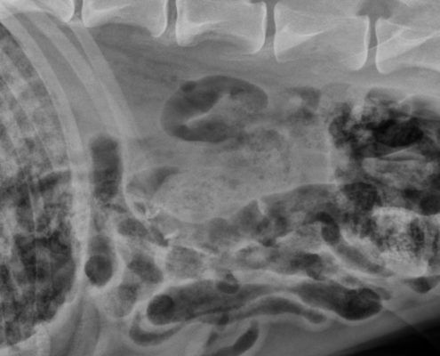
admin2017-06-27 08:00:512017-06-26 14:59:08Imaging the acute abdomen
http://blog.vetbloom.com/wp-content/uploads/2017/06/Featured-Acute-abdomen-e1498502637272.jpg
321
845
admin
http://blog.vetbloom.com/wp-content/uploads/2017/01/VetBloom-Official-Logo-Small-e1485206678262.png
admin2017-06-27 08:00:512017-06-26 14:59:08Imaging the acute abdomen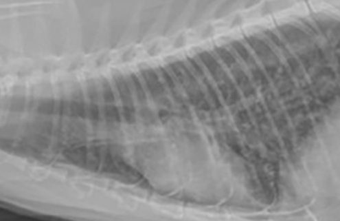 http://blog.vetbloom.com/wp-content/uploads/2017/04/Brisson-bronchial.jpg
321
845
admin
http://blog.vetbloom.com/wp-content/uploads/2017/01/VetBloom-Official-Logo-Small-e1485206678262.png
admin2017-04-18 08:00:362017-04-16 17:18:09Imaging diagnosis of common gastrointestinal and respiratory emergencies
http://blog.vetbloom.com/wp-content/uploads/2017/04/Brisson-bronchial.jpg
321
845
admin
http://blog.vetbloom.com/wp-content/uploads/2017/01/VetBloom-Official-Logo-Small-e1485206678262.png
admin2017-04-18 08:00:362017-04-16 17:18:09Imaging diagnosis of common gastrointestinal and respiratory emergencies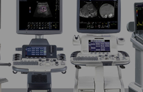 http://blog.vetbloom.com/wp-content/uploads/2016/11/Featured-Ultrasound-machines-e1510341641459.jpg
319
846
admin
http://blog.vetbloom.com/wp-content/uploads/2017/01/VetBloom-Official-Logo-Small-e1485206678262.png
admin2016-11-29 08:00:142017-03-28 09:12:49Making the most of your ultrasound machine
http://blog.vetbloom.com/wp-content/uploads/2016/11/Featured-Ultrasound-machines-e1510341641459.jpg
319
846
admin
http://blog.vetbloom.com/wp-content/uploads/2017/01/VetBloom-Official-Logo-Small-e1485206678262.png
admin2016-11-29 08:00:142017-03-28 09:12:49Making the most of your ultrasound machine http://blog.vetbloom.com/wp-content/uploads/2016/11/Featured-feline-skull-rad-e1510341697401.jpg
319
846
admin
http://blog.vetbloom.com/wp-content/uploads/2017/01/VetBloom-Official-Logo-Small-e1485206678262.png
admin2016-11-08 08:00:042016-11-07 08:55:26Feline head imaging: Continuity with practitioner and radiology
http://blog.vetbloom.com/wp-content/uploads/2016/11/Featured-feline-skull-rad-e1510341697401.jpg
319
846
admin
http://blog.vetbloom.com/wp-content/uploads/2017/01/VetBloom-Official-Logo-Small-e1485206678262.png
admin2016-11-08 08:00:042016-11-07 08:55:26Feline head imaging: Continuity with practitioner and radiology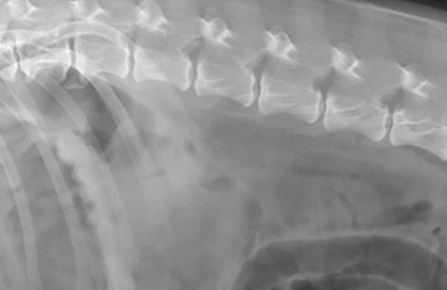 http://blog.vetbloom.com/wp-content/uploads/2016/09/Featured-pneumoperitoneum-e1510342126781.jpg
321
846
admin
http://blog.vetbloom.com/wp-content/uploads/2017/01/VetBloom-Official-Logo-Small-e1485206678262.png
admin2016-09-20 08:00:412016-09-20 07:42:26Radiography vs. ultrasound in the dog with acute abdominal signs
http://blog.vetbloom.com/wp-content/uploads/2016/09/Featured-pneumoperitoneum-e1510342126781.jpg
321
846
admin
http://blog.vetbloom.com/wp-content/uploads/2017/01/VetBloom-Official-Logo-Small-e1485206678262.png
admin2016-09-20 08:00:412016-09-20 07:42:26Radiography vs. ultrasound in the dog with acute abdominal signs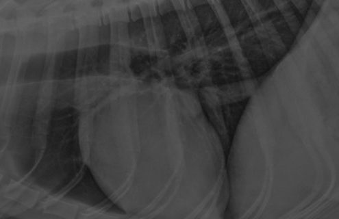 http://blog.vetbloom.com/wp-content/uploads/2016/07/Featured-teleradiology-tips-e1495535616943.jpg
320
844
admin
http://blog.vetbloom.com/wp-content/uploads/2017/01/VetBloom-Official-Logo-Small-e1485206678262.png
admin2016-07-08 08:00:072016-07-08 05:55:03Tips for digital & teleradiology
http://blog.vetbloom.com/wp-content/uploads/2016/07/Featured-teleradiology-tips-e1495535616943.jpg
320
844
admin
http://blog.vetbloom.com/wp-content/uploads/2017/01/VetBloom-Official-Logo-Small-e1485206678262.png
admin2016-07-08 08:00:072016-07-08 05:55:03Tips for digital & teleradiology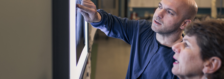 http://blog.vetbloom.com/wp-content/uploads/2015/10/25.jpg
320
900
admin
http://blog.vetbloom.com/wp-content/uploads/2017/01/VetBloom-Official-Logo-Small-e1485206678262.png
admin2016-06-14 08:00:352016-06-14 07:56:33What’s your radiographic diagnosis? Lame Maine Coon
http://blog.vetbloom.com/wp-content/uploads/2015/10/25.jpg
320
900
admin
http://blog.vetbloom.com/wp-content/uploads/2017/01/VetBloom-Official-Logo-Small-e1485206678262.png
admin2016-06-14 08:00:352016-06-14 07:56:33What’s your radiographic diagnosis? Lame Maine Coon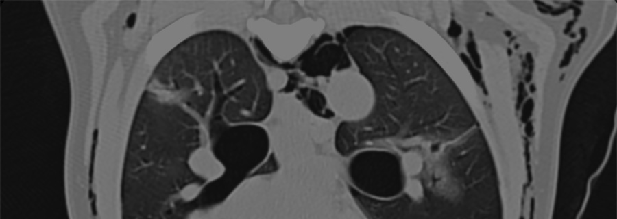 http://blog.vetbloom.com/wp-content/uploads/2016/05/Featured-Throracic-CT.jpg
320
900
admin
http://blog.vetbloom.com/wp-content/uploads/2017/01/VetBloom-Official-Logo-Small-e1485206678262.png
admin2016-05-25 16:36:532016-05-25 16:36:53Thoracic CT: More than just a pretty picture
http://blog.vetbloom.com/wp-content/uploads/2016/05/Featured-Throracic-CT.jpg
320
900
admin
http://blog.vetbloom.com/wp-content/uploads/2017/01/VetBloom-Official-Logo-Small-e1485206678262.png
admin2016-05-25 16:36:532016-05-25 16:36:53Thoracic CT: More than just a pretty picture http://blog.vetbloom.com/wp-content/uploads/2015/10/Featured-radiologists-e1461617119343.jpg
320
900
admin
http://blog.vetbloom.com/wp-content/uploads/2017/01/VetBloom-Official-Logo-Small-e1485206678262.png
admin2016-04-26 08:00:512016-04-28 14:41:50What's your radiographic diagnosis? Respiratory arrest during dental cleaning
http://blog.vetbloom.com/wp-content/uploads/2015/10/Featured-radiologists-e1461617119343.jpg
320
900
admin
http://blog.vetbloom.com/wp-content/uploads/2017/01/VetBloom-Official-Logo-Small-e1485206678262.png
admin2016-04-26 08:00:512016-04-28 14:41:50What's your radiographic diagnosis? Respiratory arrest during dental cleaning http://blog.vetbloom.com/wp-content/uploads/2015/12/Featured-Lab-puppy.jpg
320
900
admin
http://blog.vetbloom.com/wp-content/uploads/2017/01/VetBloom-Official-Logo-Small-e1485206678262.png
admin2015-12-15 08:00:032015-12-13 08:48:58Seeing between the lines
http://blog.vetbloom.com/wp-content/uploads/2015/12/Featured-Lab-puppy.jpg
320
900
admin
http://blog.vetbloom.com/wp-content/uploads/2017/01/VetBloom-Official-Logo-Small-e1485206678262.png
admin2015-12-15 08:00:032015-12-13 08:48:58Seeing between the lines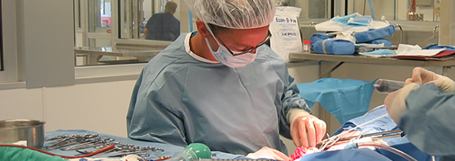 http://blog.vetbloom.com/wp-content/uploads/2015/11/Featured-surgeon.jpg
320
900
admin
http://blog.vetbloom.com/wp-content/uploads/2017/01/VetBloom-Official-Logo-Small-e1485206678262.png
admin2015-11-05 08:00:062015-11-10 14:42:12Portosystemic shunts
http://blog.vetbloom.com/wp-content/uploads/2015/10/Featured-radiologists-e1461617119343.jpg
320
900
admin
http://blog.vetbloom.com/wp-content/uploads/2017/01/VetBloom-Official-Logo-Small-e1485206678262.png
admin2015-10-27 15:02:052015-11-10 14:47:05Imaging diagnosis: Coughing German Shepherd
http://blog.vetbloom.com/wp-content/uploads/2015/11/Featured-surgeon.jpg
320
900
admin
http://blog.vetbloom.com/wp-content/uploads/2017/01/VetBloom-Official-Logo-Small-e1485206678262.png
admin2015-11-05 08:00:062015-11-10 14:42:12Portosystemic shunts
http://blog.vetbloom.com/wp-content/uploads/2015/10/Featured-radiologists-e1461617119343.jpg
320
900
admin
http://blog.vetbloom.com/wp-content/uploads/2017/01/VetBloom-Official-Logo-Small-e1485206678262.png
admin2015-10-27 15:02:052015-11-10 14:47:05Imaging diagnosis: Coughing German Shepherd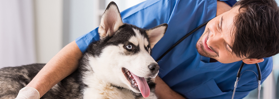 http://blog.vetbloom.com/wp-content/uploads/2015/09/5.jpg
320
900
admin
http://blog.vetbloom.com/wp-content/uploads/2017/01/VetBloom-Official-Logo-Small-e1485206678262.png
admin2015-09-10 14:20:532015-11-10 14:48:30What's your radiographic diagnosis? Vomiting Bulldog
http://blog.vetbloom.com/wp-content/uploads/2015/09/5.jpg
320
900
admin
http://blog.vetbloom.com/wp-content/uploads/2017/01/VetBloom-Official-Logo-Small-e1485206678262.png
admin2015-09-10 14:20:532015-11-10 14:48:30What's your radiographic diagnosis? Vomiting Bulldog