Minimally invasive surgery – How small can we go?
Matt Cleveland, DVM, DACVS
Veterinary Specialty Hospital by Ethos, San Diego, CA
Posted on 2016-11-01
With advances in human and veterinary medicine, new and innovative surgical techniques are constantly being devised, tested, and either discarded or adopted as the new standard. In the 1980s, human minimally invasive surgery took a big step forward with cholecystectomies completed via laparoscopy that resulted in improved patient care. With this new standard established, advances in other minimally invasive surgeries have taken hold, resulting in extension to the veterinary field. Minimally invasive procedures, like laparoscopy and thoracoscopy, have been shown to decrease patient perioperative morbidity, hospitalization time, and perioperative pain, while allowing excellent visualization and sufficient workable space to accomplish exploration, organ biopsies, organ excisions, and many other surgical procedures that have traditionally been performed in a standard open manner.
Uses in the veterinary field began with hepatic biopsies and assisted ovariohysterectomies, leading the way for subsequent expansion to more advanced minimally invasive procedures. Minimally invasive surgery has become the gold standard for certain procedures. This has resulted in adoption of these techniques as standard training protocols all surgeons must undergo to gain surgery credentialing.
Currently minimally invasive procedures that can be completed via abdominal laparoscopic procedures include ovariectomy, ovariohysterectomy, assisted gastropexy, hepatic biopsies, small intestinal biopsies, renal biopsies, adrenalectomies, splenectomies, cisterna chyli ablation, assisted cystotomies, and exploratory laparoscopy for small intestinal foreign bodies. Thoracoscopic procedures include pericardial window or subtotal pericardiectomy, thoracic duct ligation, lung biopsies or lung lobectomies, and lymph node biopsies. These advanced procedures are now more readily possible due to improvements in preoperative imaging of our patients with ultrasound, computed tomography (CT), and magnetic resonance imaging (MRI). The advanced imaging has allowed for better initial identification and classification of organ pathological changes prior to surgery. Better preoperative screening allows for a more directed surgical approach that laparoscopy and thoracoscopy can provide while minimizing incision size, increasing visceral access, and decreasing the risk of overlooking co-morbidities during the procedure. However, for the majority of these procedures, there are limitations. These depend on organ/mass size, pathologic changes, and concurrent disease processes that could make attempting the minimally invasively procedure contraindicated, all of which can be initially determined via the appropriate preoperative advanced imaging.
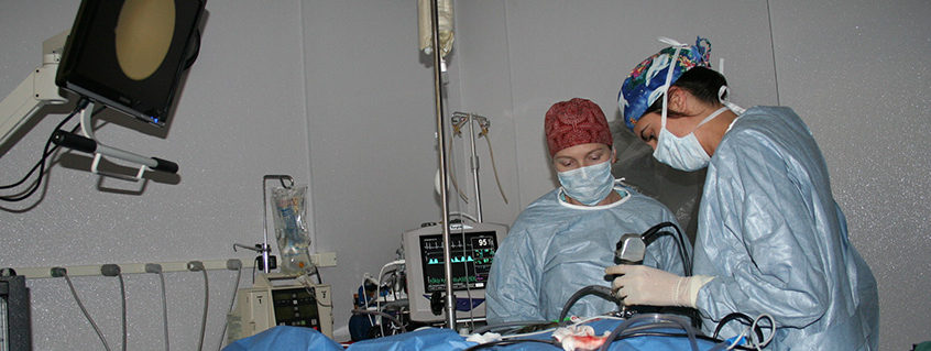
There have been multiple reports in the veterinary literature comparing laparoscopic and open procedures that document improved overall patient outcome with minimally invasive procedures. More specifically, pain scores and hospitalization times have been significantly reduced, and overall patient morbidity decreased with laparoscopic procedures when compared to open surgeries. Mainly, research has focused on ovariectomy and ovariohysterectomy, but one could easily translate those findings to other abdominal and thoracoscopic procedures. One of the great benefits is the incredibly enhanced visualization provided by the high definition cameras. This visualization allows for easier identification of visceral vascular supply. Instrumentation used in minimally invasive procedures, have enabled better hemostasis, thus reducing the risk of intra- operative hemorrhage. Newer designs of ligating device hand pieces have been created specifically for laparoscopic and thoracoscopic procedures with longer working arms and blunted (dolphin tip) nose pieces allowing for dual use of blunt dissection with quick conversion to vascular sealing and dissection. Additionally, port apertures have improved designs allowing for reduction in the quantity of ports needed. For example, the SILS port (single incision laparoscopic surgery) by Covidien allows for multiple instruments and camera devices to be used at the same time through a single, small incision. These advances have yielded an increase in the procedures which can be performed laparoscopically while continuing to reduce incision size, patient perioperative morbidity, and postoperative pain. An additional feature of minimally invasive procedures, like thoracoscopy and laparoscopy, is the simplicity in converting to a standard open approach if complications arise or if the completion of the procedure is becoming difficult. In general, complications and/or the need for conversion to open approaches are rare, especially in the hands of trained surgeons.
Laparoscopy and thoracoscopy have become the new standard of care for specific procedures as we as veterinarians strive to provide our patients with better operative care. Decreasing perioperative pain, hospitalization time, and incision size are just a few of the documented benefits that minimally invasive procedures provide. In the hands of trained surgeons, more advanced procedures like adrenalectomy and thoracic duct ligation are readily possible and should be considered for select patients. Many of these have been performed at VSH for several years. As we continue to improve preoperative imaging, intraoperative instrumentation, and optimize postoperative pain management, we will hopefully see minimally invasive procedures become the gold standard for our small animal patients requiring surgical intervention.
About the author
Dr. Cleveland is a graduate of the St. George’s University School of Veterinary Medicine, completing his clinical 4th year training at Texas A&M (2010). Following graduation, Dr. Cleveland completed a small animal rotating internship with the Pittsburgh Veterinary Specialty and Emergency Hospital and a specialty surgical internship here with the Veterinary Specialty Hospital of San Diego. Dr. Cleveland was then accepted into a three year small animal surgical residency with Angell Animal Medical Center in Boston MA, completed in 2015. He became a board-certified veterinary surgeon in March, 2016. In his free time, Dr. Cleveland enjoys surfing, fishing, skiing, and hiking, but mostly he loves spending time with his spouse Elizabeth, dogs, Madelyn and Archer, and cat, Dizzy. |