Calcium oxalate urolithiasis
Rachel Cooper, DVM, DACVIM
Massachusetts Veterinary Referal Hospital
Posted on 5 April 2016
Urolithiasis refers to the formation of stones anywhere within the upper and lower urinary tracts. It is a common problem among both canine and feline patients in veterinary medicine and one of the most common causes of lower urinary tract signs.
Urolithiasis is not a disease entity in itself, but rather a clinical consequence of several different disease entities. Determining the make-up of the urolith will help give clues into the causes, treatment and prevention of further urolith formation. There are no protocols to help promote dissolution of calcium oxalate uroliths so they have to be physically removed.
In a recent review of all stones submitted to the Minnesota Urolith Center, Calcium Oxalate stones represented 42% of all stones submitted. It is now the second most common cause of stones in both dogs and cats. Dogs that are affected with calcium oxalate urolithiasis are typically middle aged to older male small breed dogs. Affected cats are typically middle-aged to older male or female cats.
Causes
The cause for formation of calcium oxalate urolithiasis is incompletely understood and may be multifactorial. Both hypercalciuria and hyperoxaluria can predispose to calcium oxalate stone formation. Hypercalcemia promotes hypercalciuria which may lead to formation of calcium oxalate stones. Approximately 35% of cats and 4% of dogs with calcium oxalate urolithiasis were hypercalcemic. Ionized calcium should be always be checked in animals with calcium oxalate urolithiasis for elevations, even with a normal total calcium. In one study of cats with hypercalcemia, 25% had normal total calcium and only ionized hypercalcemia. The most common cause of hypercalcemia in cats with calcium oxalate urolithiasis was idiopathic hypercalcemia, while the most common cause in dogs was primary hyperparathyroidism. Other causes of hypercalciuria could include excessive dietary calcium, animal protein and/or vitamin D consumption, medications that cause calcium elimination in the urine (furosemide, prednisone), vitamin B6 deficiency, hyperadrenocorticism, metabolic acidosis and excessive oxalate excretion from excessive dietary intake of oxalate and/or ascorbic acid.
Diagnosis
Radiography is the cornerstone for diagnosis of urolithiasis. Calcium oxalate uroliths are radioopaque, so they are easily seen on abdominal radiograph when greater than 2-3mm in diameter. Pneumocystography (negative contrast cystography) is more sensitive at detecting bladder stones than regular radiography, with a false negative rate of 6.5%. Adding a small amount of contrast and performing a double-contrast cystography improves accuracy with a false negative rate of 5%. Ultrasound is as sensitive as double contrast radiography for detection of bladder stones but is not useful for further characterization of number and/or size of stones.
Treatment
Voiding urohydropulsion is a technique that can be used to evacuate uroliths of a small to moderate size (<5-7mm in diameter, depending on the size and sex of the patient). The patient is placed under general anesthesia and a catheter is advanced through the urethra into the bladder. Sterile saline is injected into the catheter to distend the bladder. The degree of bladder distension is assessed by abdominal palpation. Once the bladder is appropriately distended, the patient is repositioned so the spine is approximately vertical. This will cause the uroliths to fall into the trigone of the bladder. The bladder is agitated at this time to facilitate movement of the stones. Steady constant pressure is applied to the bladder to induce urination, and once urination starts, the bladder is more vigorously expressed until it is empty. The stream of urine can be caught and assessed for stone expulsion. This process may be repeated until all stones are flushed out. Double contrast cystography is advised at the end of this procedure to ensure no small stones are left.
Voiding urohydropulsion can be used as a sole therapy for removal, or can be used in addition to other therapies. This procedure is generally very safe when the correct technique is performed in a good candidate. The most common complications of voiding urohydropulsion is visible hematuria. Urethral obstruction with uroliths can occur during voiding urohydropulsion. If this should occur, the ureterolith should be flushed back into the bladder and removed via cystotomy or cystoscopy, if possible. Ruptured bladder is an uncommon risk of voiding urohydropulsion. This procedure should not be used in dogs or cats with urinary tract infections, urethral obstructions, or history of recent bladder surgery. This procedure can be performed as a sole therapy, or in conjunction with cystoscopy +/- lithotripsy.
Cystoscopy +/- laser lithotripsy can also be used for stone removal. Cystoscopy is performed with the patient under general anesthesia. This procedure is ideal for a single to a few larger stones. Once the stones are visualized, a basket is passed under cystoscopic guidance. The basket is placed around the stone, and then the entire apparatus is removed. This procedure is most useful in female cats for stones 3-5mm and female dogs for stones 4mm-1cm varying on the size of the dog. In male dogs, due to the small diameter and longer length of their urethras, this can be attempted for few stones 2-3mm in diameter. For stones that are larger than this, laser lithotripsy can be performed under cystoscopic guidance and the fragments can be removed via stone baskets with cystoscopy, or via voiding urohydropulsion.
Percutaneous cystolithotomy is a new technique used to remove stones from the bladders of dogs and cats. This method is performed via a small ventral midline skin incision made directly over the bladder. A trocar is advanced into the bladder lumen and a rigid cystoscope is advanced through the trocar into the urinary bladder. Stone removal is performed with an endoscopic stone basket. The advantages of this technique are that the entire mucosal surface of the bladder and the urethra can be evaluated with high powered magnification of the cystoscope. After the stones are removed the bladder is closed and the skin incision is closed. This procedure can be performed on an outpatient basis. A recent article (J Am Vet Med Assoc 2011;239:344-9) evaluated the use of PCCL in a population of dogs and cats with cystoliths. In that population of 27 animals, the median procedure time was 66 minutes, and no complications were noted.
Routine cystotomy is also a reasonable alternative for stone removal, especially with a large number of stones or multiple very large stones. In a recent study (J Am Vet Med Assoc 2010;236:763-6), 20% of dogs had incomplete removal of bladder stones via cystotomy. Performing post-operative radiographs (ideally a double contrast cystogram) is considered a standard of care to help prevent this occurrence.
Prevention
Risk factors that promote hypercalciuria and oxaluria should be addressed.
Since urolith recurrence is very common, follow-up radiographs every 2-3 months for the first year, then every 3-6 months thereafter to look for recurrence of stones. If small stones are found early, they can be removed by voiding urohydropulsion and prevent the need for an additional surgical procedure.
Increased water intake is essential to decrease the risk of recurrent urolithiasis. This can be accomplished by feeding a canned food diet and encouraging water consumption. Urinalysis should be monitored for urine specific gravity, calcium oxalate or struvite crystalluria and/or signs of inflammation every 3-6 months. Ideally, urine pH should be maintained between 6.8 and 7.2 and urine specific gravity should be less than 1.020.
Diets for dogs with calcium oxalate uroliths should contain citrate as well as have adequate phosphate and magnesium. Commercial diets that have most frequently been advocated for prevention of calcium oxalate urolithiasis include Royal Canin SO and Hill’s Prescription Diet u/d. Diets with a high fat content such as u/d should be avoided in dogs with a history of pancreatitis, obesity, diabetes mellitus or hyperlipidemia. Alternative diets that are lower in fat include Hill’s w/d or Hill’s g/d. These low-fat diets should be supplemented with oral potassium citrate. Excessive vitamin C and vitamin D intake should be avoided. Vitamin C can be broken down by the body into oxalates, increasing the risk for calcium oxalate stones. Vitamin D will increase the risk of calcium oxalate stones by increasing the excretion of calcium into the urine.
Deficiency of Vitamin B6 promotes endogenous production and excretion of oxalates, so B6 supplementation may also be advocated in these cases, especially if a home-made diet is used. For dogs with a history of calcium oxalate stones, potassium citrate (75-100mg/kg BID) may be helpful to minimize oxalate excretion. Thiazide diuretics have also been shown to lower calcium oxalate supersaturation in the urine and theoretically can be used to decrease the likelihood of calcium oxalate stone formation if the above therapies are not effective. Thiazide diuretics promote calcium retention, so they are contraindicated if hypercalcemia is a contributing factor.
New therapies that are currently being investigated for prevention of calcium oxalate urolithiasis include probiotics. Oxalobacter formigenes is a non-pathogenic intestinal anaerobic microbe that ingests oxalates as its sole nutrient. With more oxalate removed in the intestine, less oxalate is available for absorption into the bloodstream, decreasing blood and urine oxalate concentrations. O. formigenes have shown to decrease urinary oxalate levels in humans, but no clinical studies have yet been conducted in veterinary medicine. Other intestinal bacteria can also metabolize intestinal oxalate.
Calcium oxalate urolithiasis is a frustrating problem for both clients and owners, with a recurrence rate approaching 50% within three years of initial diagnosis. With measures to decrease risk of recurrence and careful monitoring, if recurrences happen they can be managed using less invasive techniques, voiding urohydropulsion, or cystoscopic guided stone retrieval.
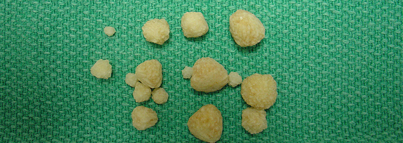
Image at top of page used with permission; contributed by Joel Mills. https://en.wikipedia.org/wiki/Bladder_stone_(animal)#/media/File:Calcium_oxalate_stones.JPG


