Tear film deficiencies: Quality vs. quantity
Emily Moeller, DVM, DACVO
Veterinary Specialty Hospital, San Diego, CA
Posted on 2018-01-09 in Ophthalmology
Keratoconjunctivitis Sicca (KCS) is one of the most commonly diagnosed diseases in veterinary ophthalmology. Common signs include conjunctival hyperemia, mucoid ocular discharge, lackluster corneal and conjunctival surfaces, and keratitis. An abundant, stable tear film is essential for the health of the cornea.
The precorneal tear film consists of three layers. The surface layer is the lipid layer which is composed of oil. It is produced by the Meibomian glands of the eyelids and prevents premature evaporation of the underlying aqueous layer. The aqueous layer is produced by the lacrimal glands of the orbit and third eyelid. The aqueous layer provides oxygen, glucose, electrolytes and water to the corneal surface. The deepest layer of the tear film is composed of mucin. Mucin is produced by the goblet cells of the conjunctiva. The mucin creates an interface between the aqueous layer and the corneal surface, which is hydrophobic. Tear film abnormalities can occur with a deficiency in any of these components. Two broad categories of tear film abnormalities are recognized: quantitative and qualitative deficiencies.
Quantitative tear deficiencies are caused by a lack of aqueous tear production. Keratoconjunctivis sicca is considered a quantitative tear film deficiency. Aqueous tear production can be measured with the Schirmer Tear Test (STT) and Phenyl Red Thread test. In cats, measurement of aqueous tear production is generally not useful because tear production in clinically normal cats is highly variable.
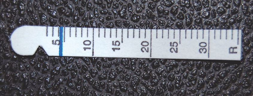
The Schirmer Tear Test can be performed in two ways. The most commonly performed is the Schirmer Tear Test I, which is performed by placing the tip of a standardized, demarcated filter paper strip into the lower conjunctival fornix for 1 minute. The STT I measures basal tear production (tears pooled on eyelid) and reflexive tear production (tearing that occurs as a reaction to the irritating strip). KCS in dogs is generally diagnosed by an STT I value of < 15mm wetting/minute. Tear production in puppies may not reach normal levels until 9-10 weeks of age. The STT II is similar to the STT I, but is performed a few minutes after application of topical anesthetic. Because the anesthetic eliminates reflexive tearing, the STT II measures basal tear production only. An STT II of > 10mm wetting/ minute is considered normal in the dog.
The Phenyl Red Thread test is performed by placing the tip of a thread impregnated with phenol red into the lower fornix for 15 seconds. This test is less irritating than the STT and can be performed in smaller species. When the phenol red comes into contact with alkaline tears, it changes color from yellow to red. With this test, normal tear production in dogs ranges from 29-38mm wetting/minute.
Qualitative tear film deficiencies refer to deficiencies in the stability of the tear film, caused by a deficiency in the lipid and/or mucin layer. Patients with qualitative tear film deficiencies typically have normal or high STT values but have ocular surface disease (ie keratitis, conjunctivitis and irritation) consistent with KCS. Lipid deficiencies can be caused by blepharitis. Mucin deficiencies are common with conjunctivitis. The tear film quality can be evaluated with the Tear Film Breakup Time (TFBUT).
The tear film breakup time is evaluated by placing a drop of fluorescein dye onto the corneal surface, holding the eyelids open, and watching (with a cobalt light source) for development of dry spots in the tear film. The dry spots will look like black defects in the surrounding fluorescein. The TFBUT is calculated as the time between application of the dye and development of the first dry spot on the cornea. A normal TFBUT is greater than 20 seconds.
Treatment for quantitative tear film deficiencies involves the use of lacrimomimetics (artificial tear solutions) as well as lacrimostimulants such as cyclosporine and tacrolimus. Treatment for a qualitative tear film deficiency is dependent on the source of the deficiency. Therapy for blepharitis (such as systemic antibiotics) and warm compressing can improve the health and availability of the meibomian gland secretions. Artificial tear ointments supplement the deficient lipid layer. For mucin deficiencies, topical anti-inflammatories to address conjunctivitis and treatment with mucinomimetics (artificial tear gels) can significantly improve clinical signs and tear film stability.
About the author
|
 Dr. Emily Moeller is a San Diego native. She attended the University of California, Berkeley with an undergraduate degree in Integrative Biology and a minor in music. She attended veterinary school at the University of California, Davis. Following veterinary school she completed a rotating internship in small animal medicine and surgery at the Veterinary Specialty Hospital. She then completed a residency in veterinary ophthalmology and was board certified as a Diplomate of the American College of Veterinary Ophthalmologists in 2011. Following residency, she practiced in Tucson, Arizona and has recently returned home to San Diego. Her special interests include ocular manifestations of systemic disease, corneal disease, and glaucoma. Her practice extends to all animal species. When at home, she enjoys cooking, playing music, and spending time with her poodle mix, Bea.

